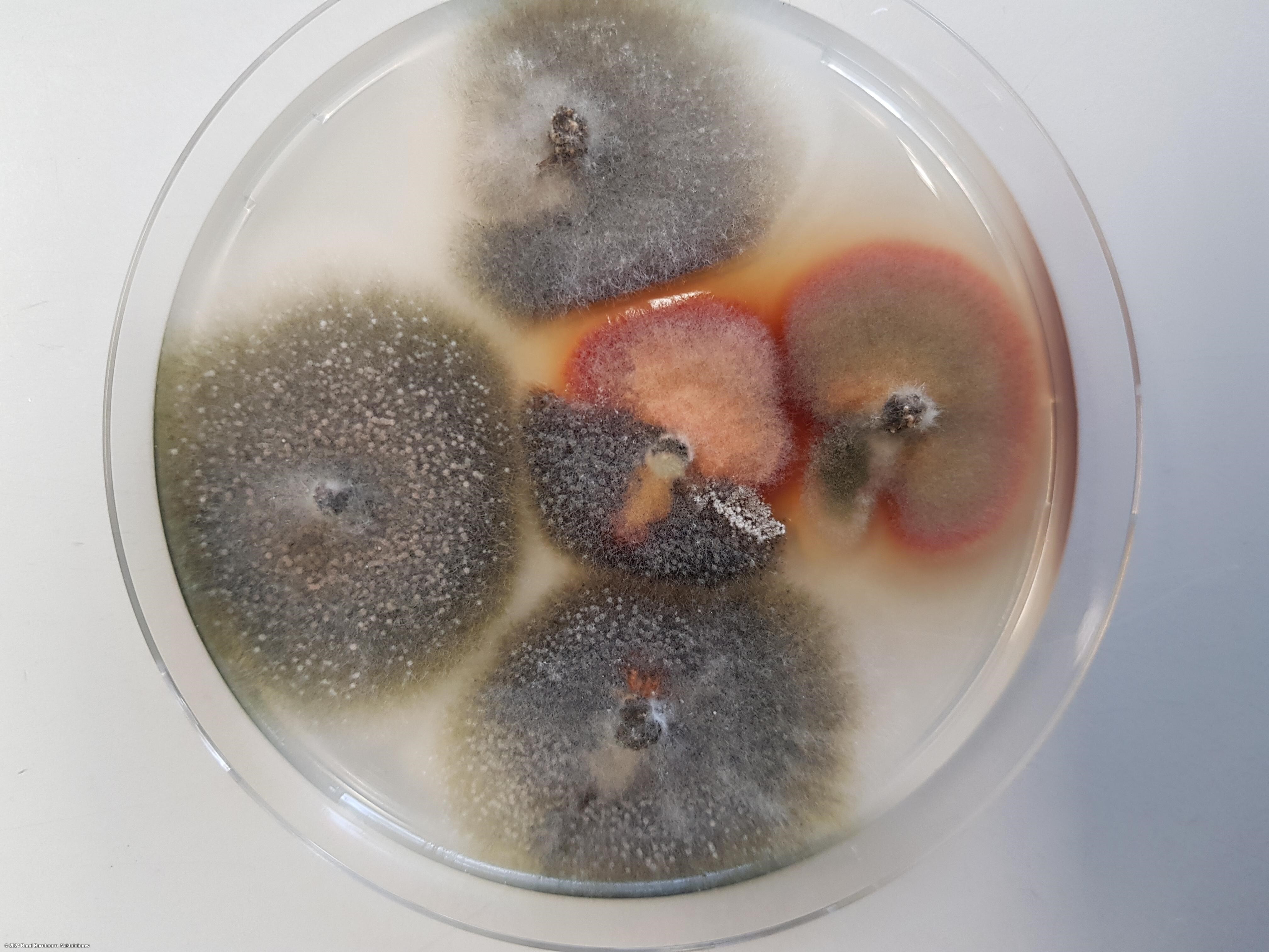
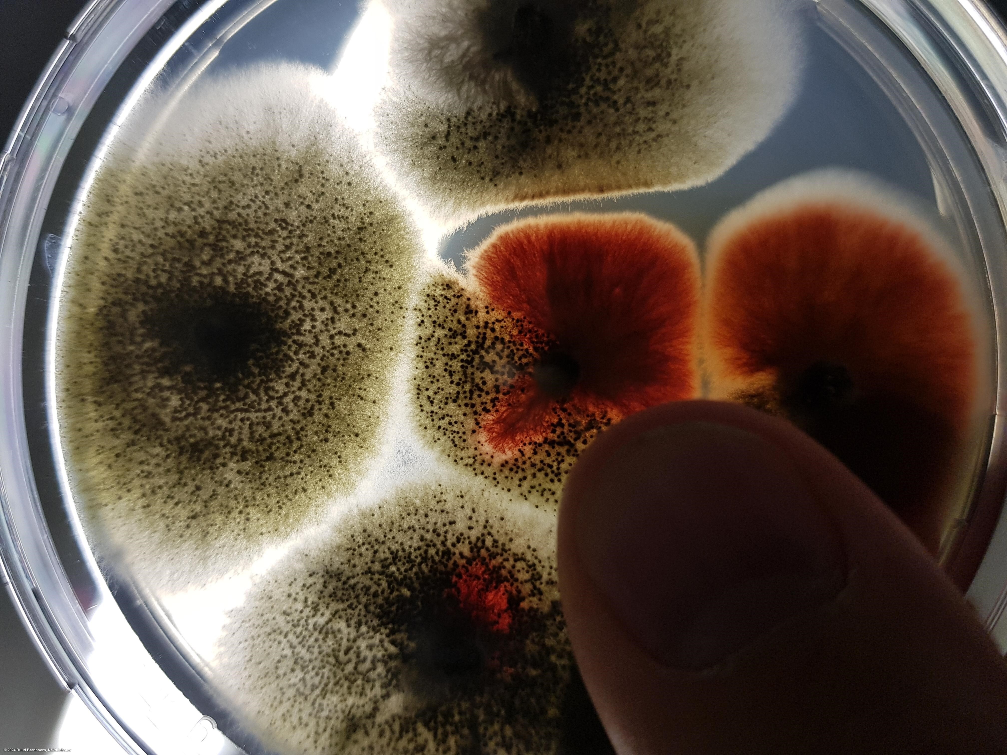
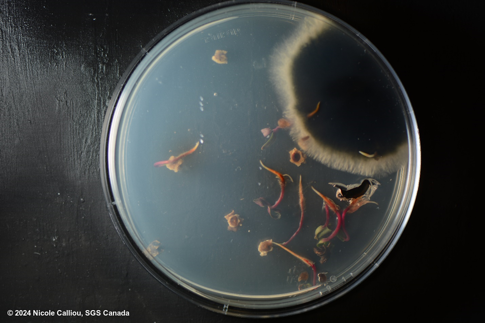
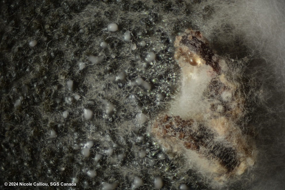
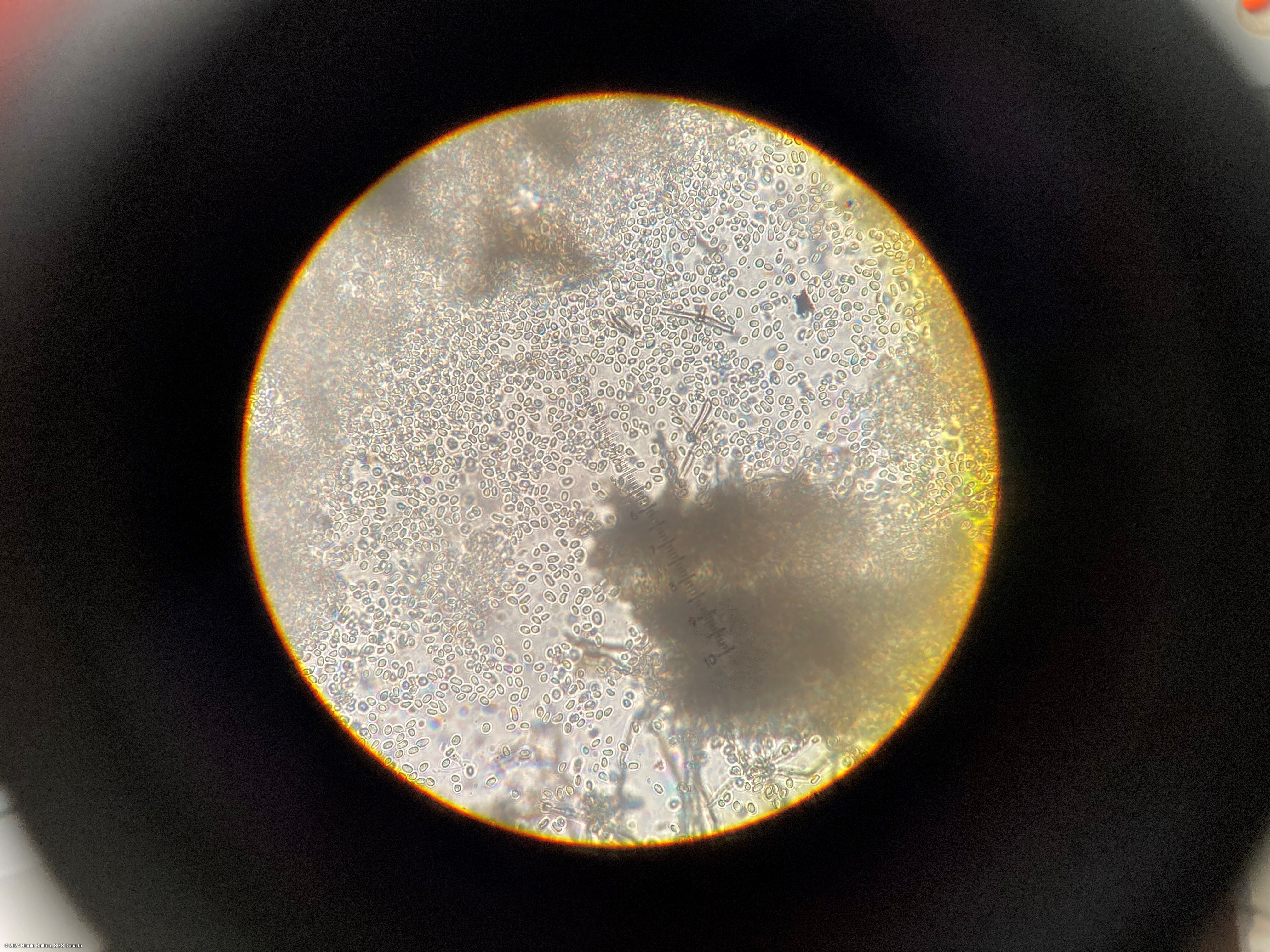
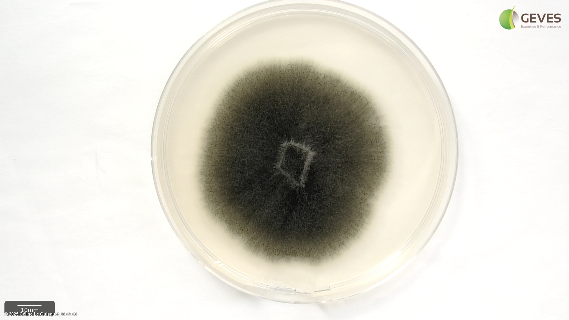
Colonies are relatively rapid growing on PDA, and show white to smokey grey coloration. Numerous black pycnidia can be seen, with obvious oozing (white) throughout the colony. Reverse/bottom of colony is significantly darker that the top of the colony (dark grey-black coloration). Pycnidiospores are hyaline, aseptate and small in size (5-8 x 2.5-4 μm) and may be extruded from the ostiole of the pycnidium.
From ISTA Working Sheet #49:
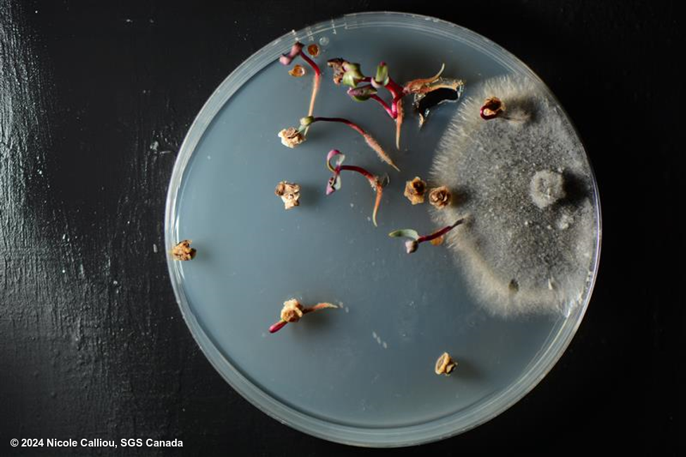
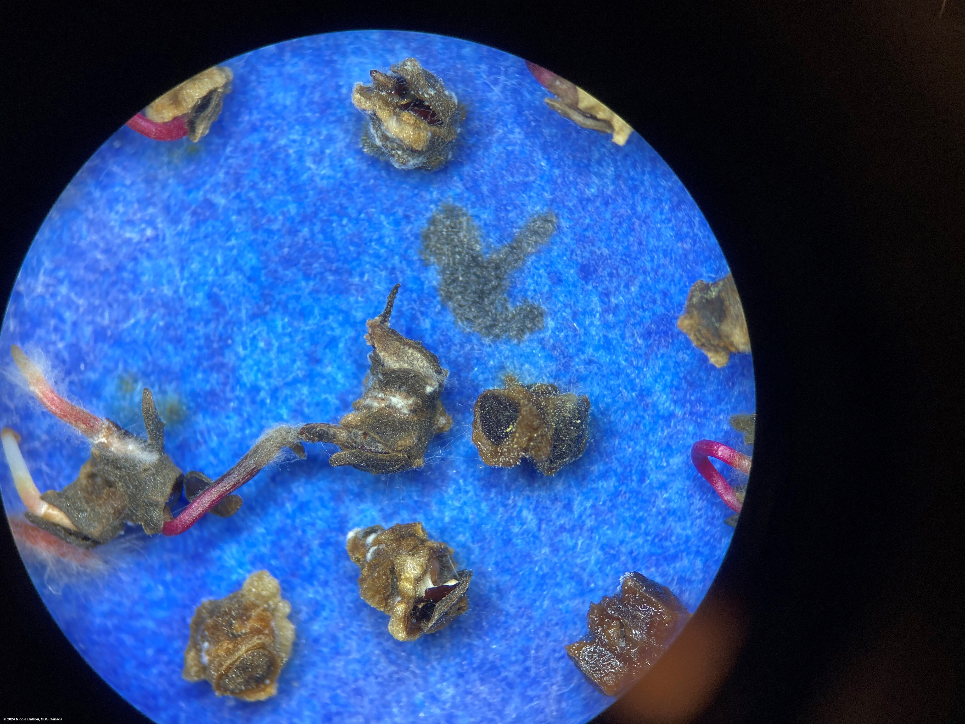
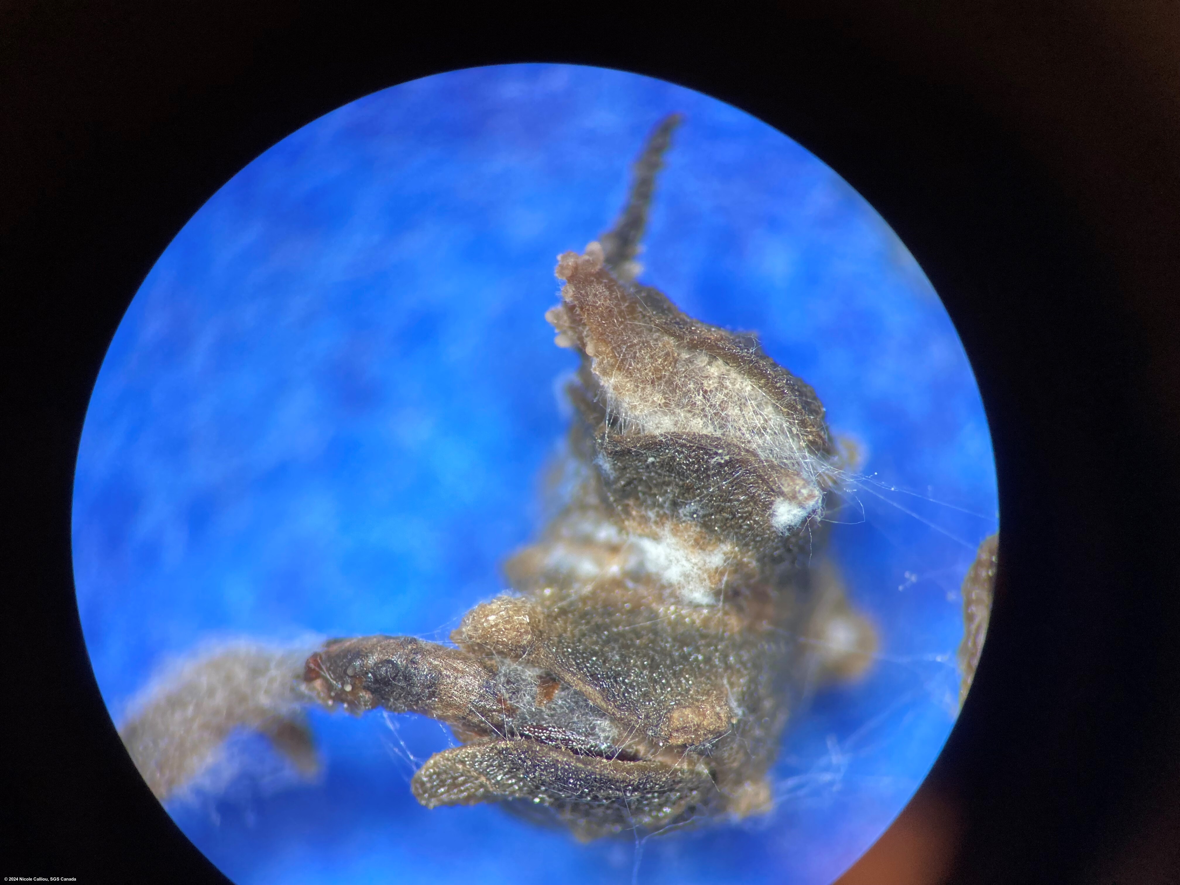
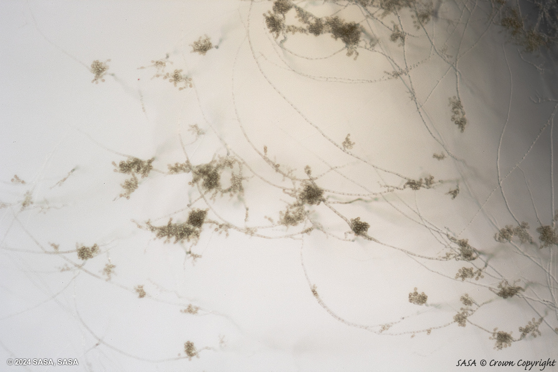
On Water agar (1.2%) with 50ppm sodium salt of 2,4-D: AFter 7 days growth at 20C in darkness, remove seeds and seedlings and examine bottom of the plate under x40-60 magnification for structures referred to as holdfasts. The holdfasts are formed only at the bottom of the dish and correct focusing is important. They are typically formed at the tips of descending, sometimes spiral, hyphae as a number of hyaline swellings. In the presence of bacteria, holdfasts may be restricted in development and are then brown in colour. Few hyphal formations of other fungi have been noted which might be confused with holdfasts. Any such structures are formed throughout the layer of agar, and not just at the bottom of the dish. This is the case with chlamydospores of Fusarium spp. which are commonly intercalary and also formed on finer hyphae than those of P. betae.
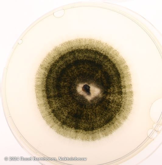
On PDA: after 7 days growth at 20C under constant nUV, examine plate for dark green colonies with pycnidia. Under constant nUV mycelial growth is kept to a minimum and pycnidia are readily visible.