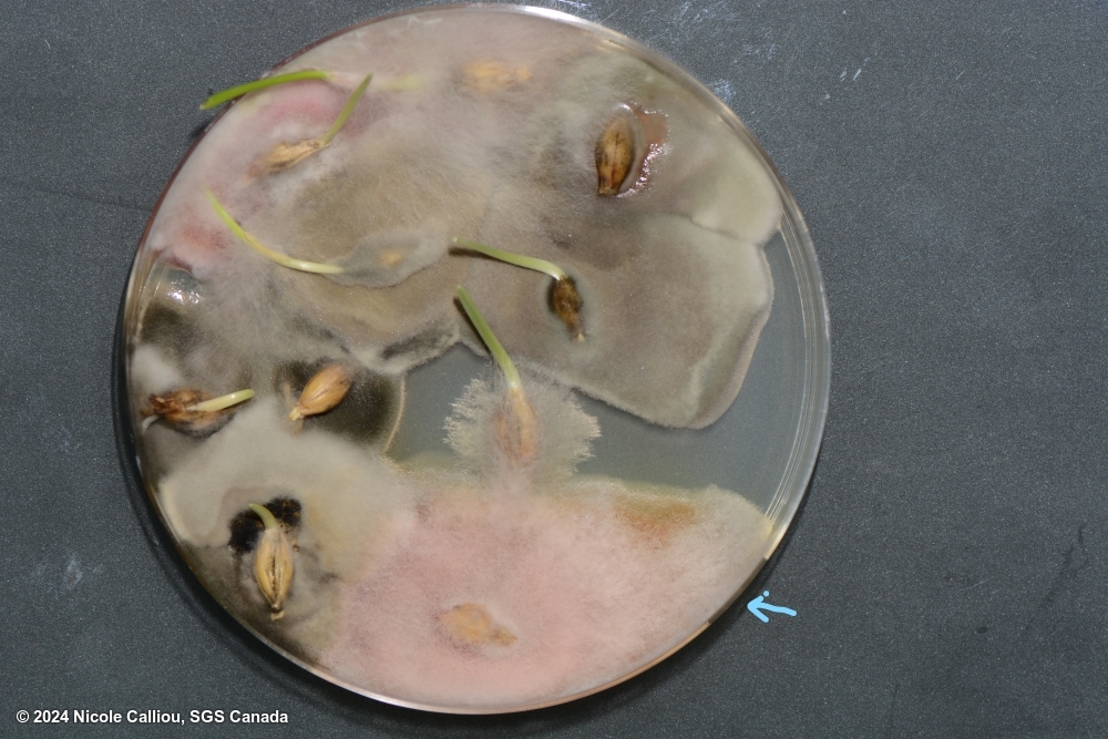
Fusarium poae on Hordeum vulgare subsp. vulgare
Image credits: Nicole Calliou, CA
Plates show both an F. poae (marked with blue arrow) and an F. graminearum colony. Spore image shows abundant microconidia, and microconidia on monophialide
sterilisation: 1% NaOCl 4 min | growth time: 5 days | temperature: 25+/-2C | lighting: 3:1 (fluorescent:nUV), within 60cm of light. Alternating light/dark in 12 hr intervals | plate size: 100mm | agar: PDA ( Neogen) |
DNA testing: None
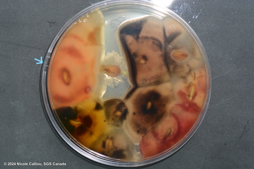
Fusarium poae on Hordeum vulgare subsp. vulgare
Image credits: Nicole Calliou, CA
Plates show both an F. poae (marked with blue arrow) and an F. graminearum colony. Spore image shows abundant microconidia, and microconidia on monophialide
sterilisation: 1% NaOCl 4 min | growth time: 5 days | temperature: 25+/-2C | lighting: 3:1 (fluorescent:nUV), within 60cm of light. Alternating light/dark in 12 hr intervals | plate size: 100mm | agar: PDA ( Neogen) |
DNA testing: None
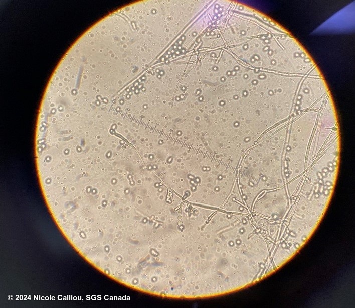
Fusarium poae on Hordeum vulgare subsp. vulgare
Image credits: Nicole Calliou, CA
Plates show both an F. poae (marked with blue arrow) and an F. graminearum colony. Spore image shows abundant microconidia, and microconidia on monophialide
sterilisation: 1% NaOCl 4 min | growth time: 5 days | temperature: 25+/-2C | lighting: 3:1 (fluorescent:nUV), within 60cm of light. Alternating light/dark in 12 hr intervals | plate size: 100mm | agar: PDA ( Neogen) |
DNA testing: None
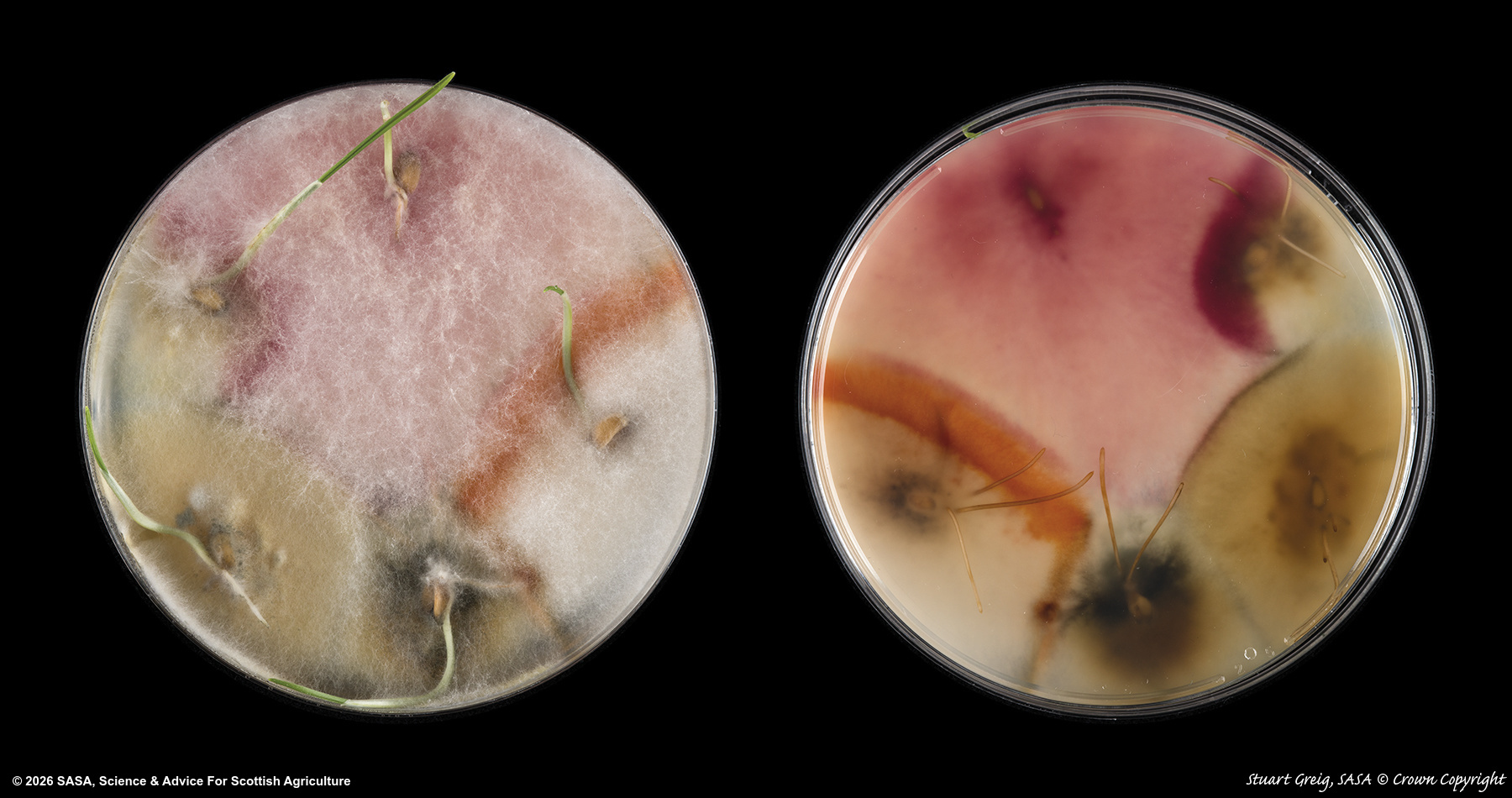
Fusarium poae on Triticum spp.
Image credits: SASA, GB
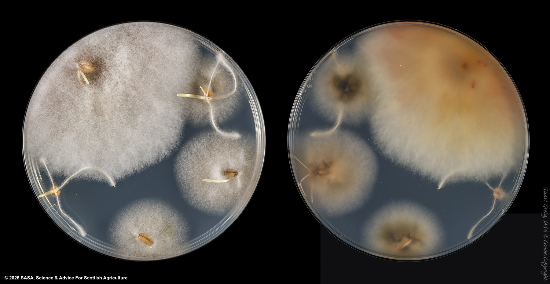
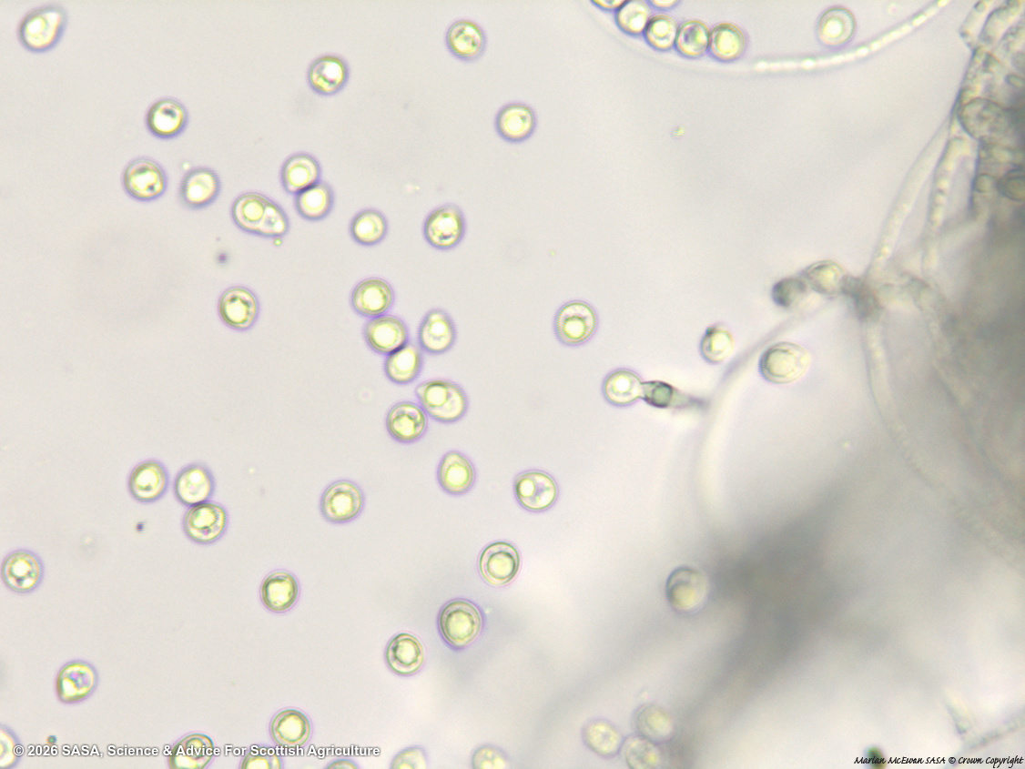
Isolates of Fusarium poae from wheat seed on Oxoid PDA, top and bottom and microconidia image with monophialide.
sterilisation: Sodium hypochlorite (1% available chlorine) 10 mins | growth time: 7 | temperature: 20C | lighting: Dark | plate size: 90mm | agar: PDA ( Oxoid)
Fusarium poae on Triticum spp.
Image credits: SASA, GB
Isolates of Fusarium poae from wheat seed on Oxoid PDA, top and bottom and microconidia image with monophialide.
sterilisation: Sodium hypochlorite (1% available chlorine) 10 mins | growth time: 7 | temperature: 20C | lighting: Dark | plate size: 90mm | agar: PDA ( Oxoid)
Fusarium poae on Triticum spp.
Image credits: SASA, GB
Isolates of Fusarium poae from wheat seed on Oxoid PDA, top and bottom and microconidia image with monophialide.
sterilisation: Sodium hypochlorite (1% available chlorine) 10 mins | growth time: 7 | temperature: 20C | lighting: Dark | plate size: 90mm | agar: PDA ( Oxoid)
Fusarium poae on Triticum spp.
Image credits: SASA, GB
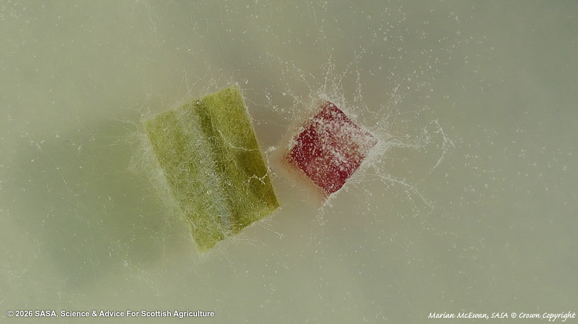
Fusarium poae plug on Carnation Leaf Agar. The F. poae plug is the pink square, and the green rectangle is part of the carnation leaf for carnation leaf agar. Please note that macroscopic photographs of Fusarium colonies grown on CLA have limited diagnostic value. CLA is mainly used to promote sporulation and facilitate the observation of microscopic structures. At the colony level, many Fusarium species appear very similar, typically showing white to cream or pale pink colonies, sparse aerial mycelium, relatively slow growth, and salmon-colored sporodochia. These macroscopic traits are often too variable and overlapping to allow reliable species identification.
growth time: 7 | temperature: 20C | lighting: 12 hours NUV | plate size: 90mm | agar: Carnation Leaf Agar ( Oxoid Technical No 3 )