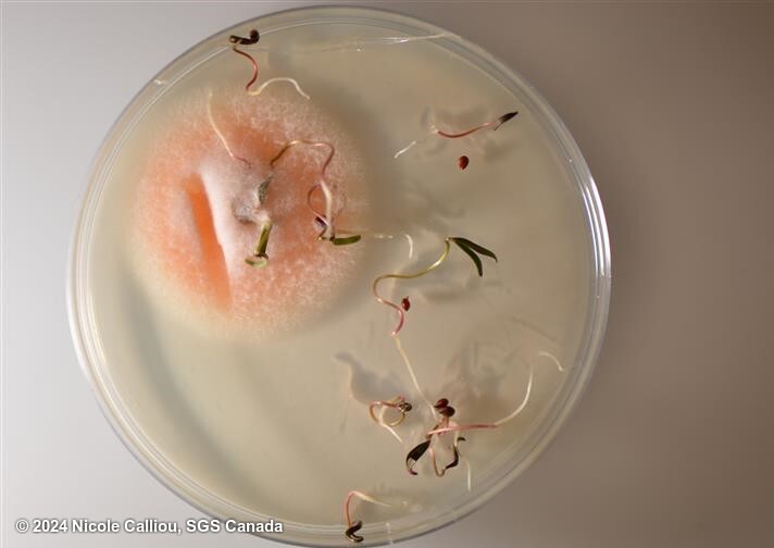
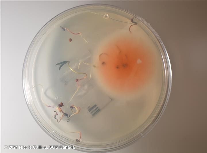
Fusarium oxysporum (Snyder and Hansen, 1940) is a soil and seed transmitted pathogen group in a large amount of different plant hosts. It considered as one of the most abundantly present fungi found in and around root systems of plants. Although the large amount of pathogenic variants are known to be present, an even larger amount of non-pathogenic variants do also exists. All these variants have the ability to penetrate the roots of plants, but only the pathogenic variants have the ability to move into the plants vascular system and, by clothing the xylem, cause wilting of plants. To accommodate the known pathogenic variants within Fusarium oxysporum, Snyder& Hansen erected 25 formae speciales (Snyder and Hansen, 1940). The formae specialis categorisation is based on pathogenicity only to a certain crop type e.x. Fusarium oxysporum formae specialis lycopersici being only pathogenic in tomato. Identification of Fusarium oxysporum is morphologically based, principally on the shape of the macroconidium, the structure of the micro conidiophore, and the formation and disposition of chlamydospores. In laboratory tests seeds are transferred to growing media and fungi looking fluffy white to orange and pink to purple are investigated via microscopy. To determine if the Fusarium oxysporum on the plate is a pathogenic variant molecular discrimination on formae specialis level via PCR is an option. Also, a more time consuming option is the use of a pathogenicity assay.
From ISTA Working Sheet #58:
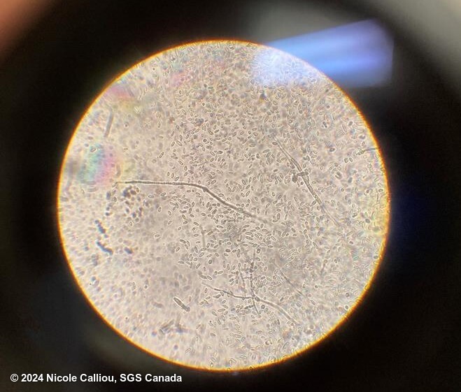
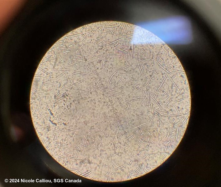
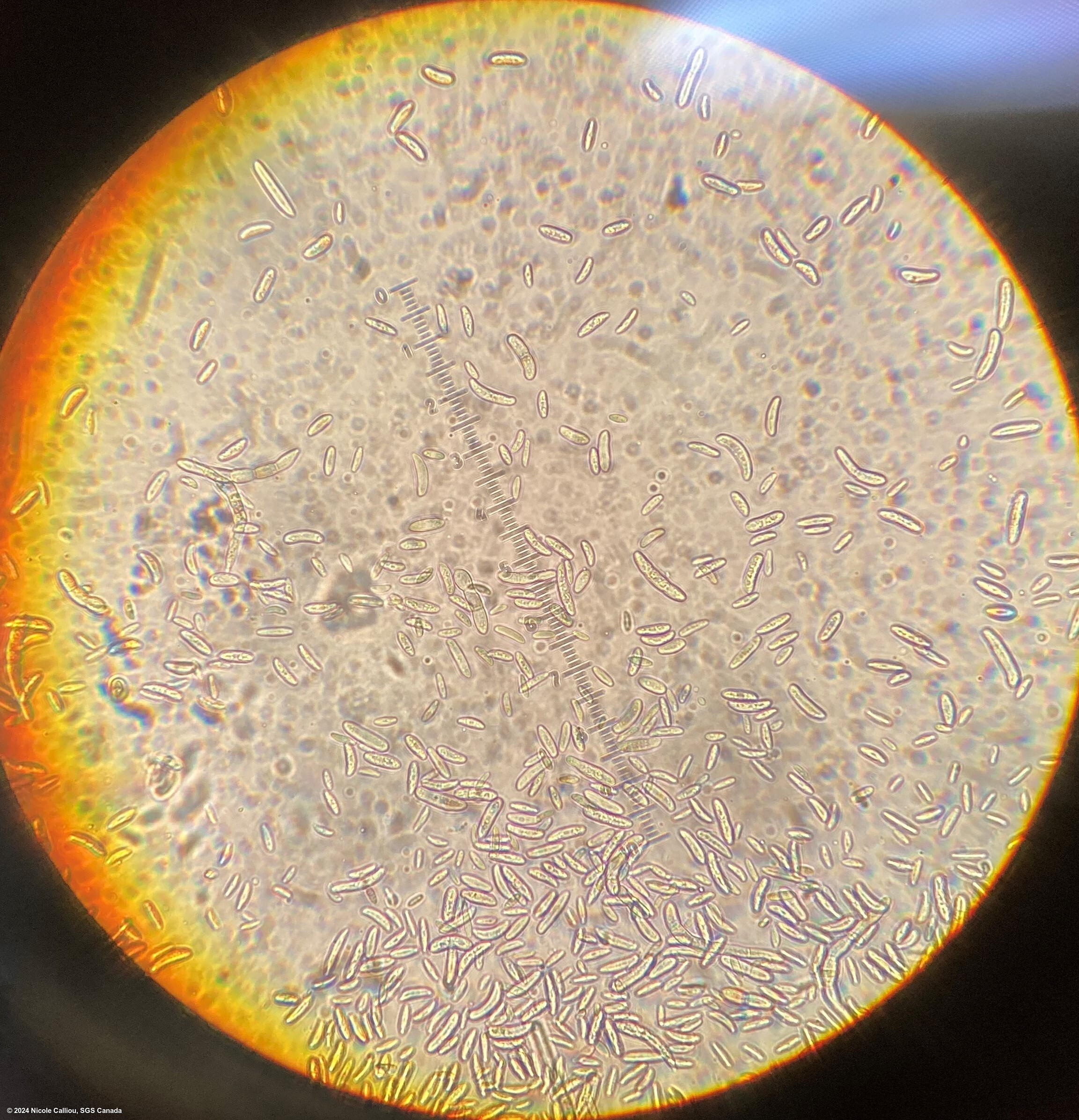
On Littman-agar, after 5 days growth at 25C with 12hr alternating dark and nUV light, examine plates with unaided eye for presence of fast growing colonies showing variable pigmentation, from pale beige to rose, purple, vinaceous, blue. Check under a stereoscopic and a compound microscope. Microconidia in false heads, mostly 1-celled, oval to reniform measuring 5-12 x 2.2-3.5 μm. Macroconidia in globose sporodochia or in pionnotal slimes, falcate, gradually tapering toward both ends, with a pointed or slightly hooked apical cell, and a foot shaped basal cell, from 0- to 7-, mostly 3-septate, ranging form 4-13 x 2.0-3.5 μm to 34-78 x 3.0-6.0 μm, predominantly 27-46 x 3-4.5 μm. Phialides monophialidic, chlamydospores globose, generally abundant, in hyphae and in conidia, terminal or intercalary, both smooth and rough walled, single, in pairs or short shains.
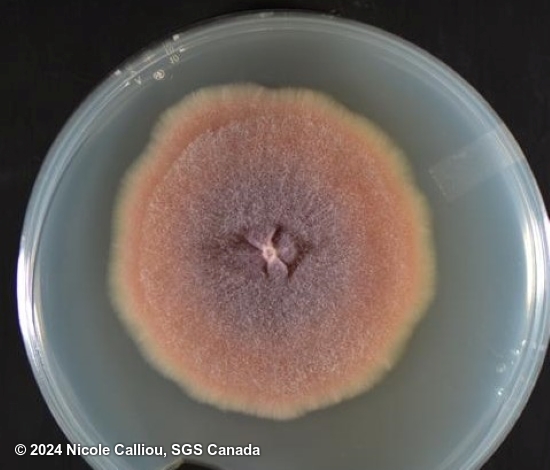
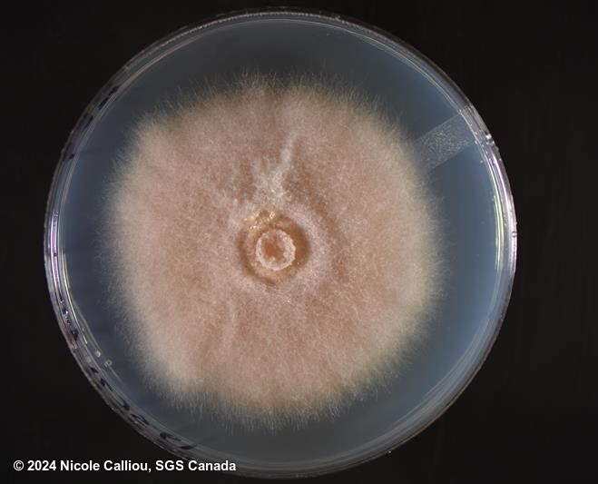
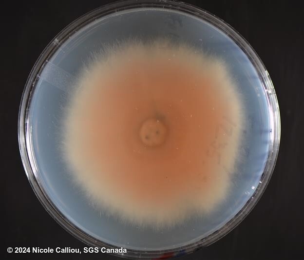
The Littman-agar is a growth restricting medium, different selective media may show more of less colour in the developing colonies. The fungus, transferred to tubes of boiled rice, produces a very characteristic colour which is alizarine pink in the lighter portions, and old rose or begonia rose in the darker portions of the colony. Isolates of the fungus may very in morphology and cultural characteristics.
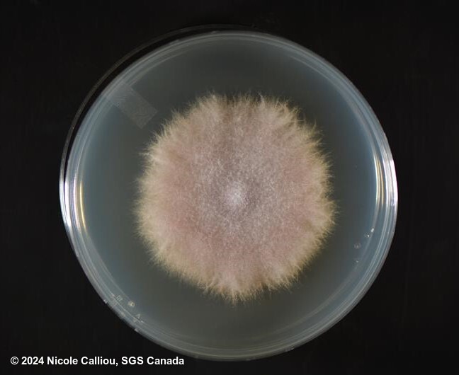
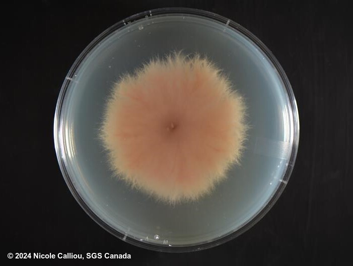
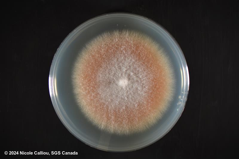
On PDA, mycelia may be floccose, sparse or abundant, and the colour range tends to be from white to pale purple. Pale orange or purple central spore masses may be present with abundant macroconidia. Dark sclerotia may be, or may not, be produced. Some isolates will readily mutate to a pionnotal form (to a flat, 'wet' mycelial colony), and then the colour may also be found to be yellow or orange.