Neocamarosporium betae
Neocamarosporium
PLEOBJ
Phoma beet spot
Phoma betae
Beta vulgaris subsp. vulgaris
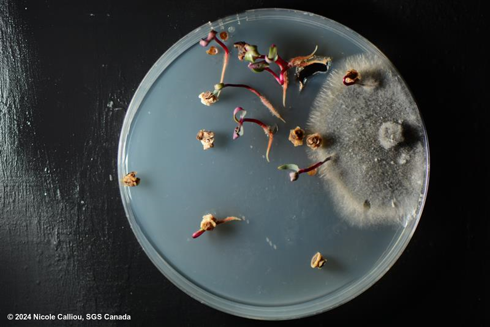
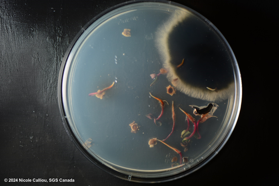
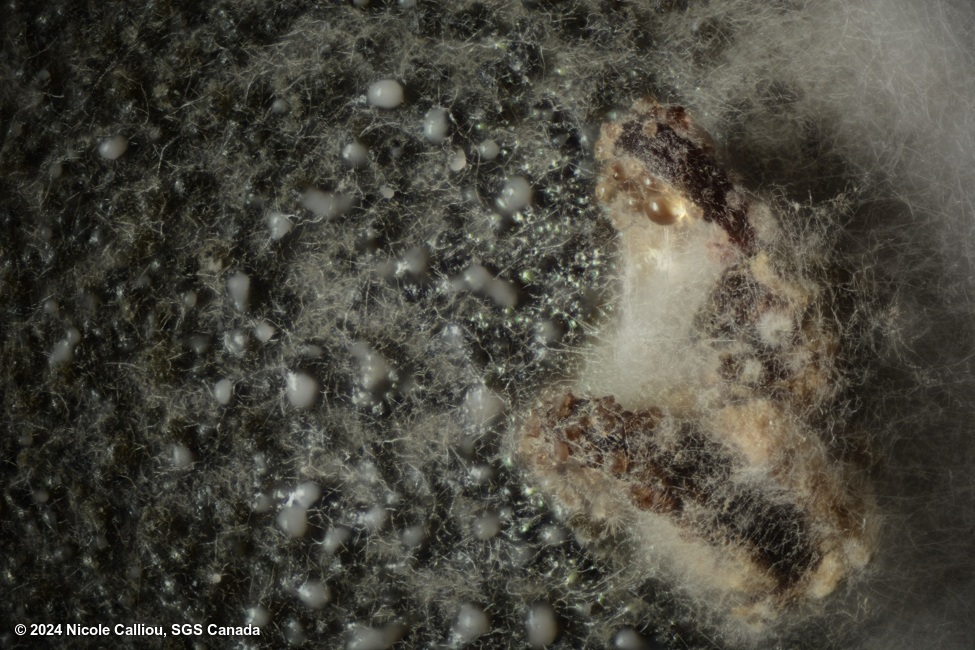
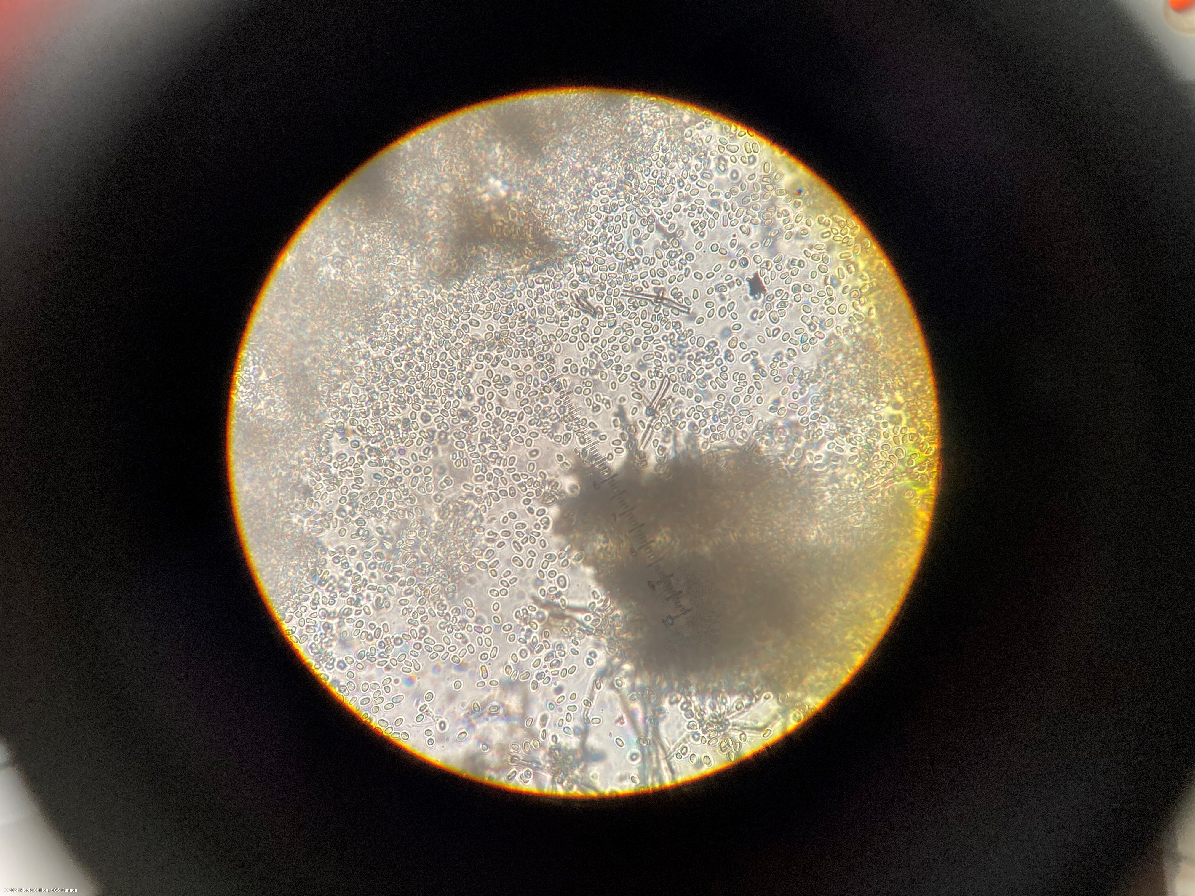
Figure 1 -2: Dark grey colony of N. betae from top and bottom. Figure 3 is the light microscope at 400x zoom showing the pycnidiospores. Figure 4 is a close up showing the oozing of the pycnidia.